Published Research
Lung Cancer Subtyping from Chest CT
Which classifier actually wins on ResNet-50 features? A measured comparison
Best Paper
Award
Natural Sciences category, KOUBAK'26
89.5%
Best accuracy
XGBoost, held-out test set
4
Classifiers compared
Overview
Lung cancer has among the highest mortality of any oncological disease, and treatment depends on more than a yes/no answer — telling adenocarcinoma, squamous cell carcinoma and large cell carcinoma apart changes what happens next. This paper asks a narrower, more answerable question: once you have deep features from a CT scan, which classifier should you put on top of them?
Published in the proceedings of KOUBAK’26, the 1st Kocaeli University Student Congress on Scientific Research, where it won Best Paper in the Natural Sciences category (Fen Bilimleri).
Approach
Rather than fine-tuning a network end to end, the pipeline splits the problem in two. A pre-trained ResNet-50 acts purely as a feature extractor, turning each CT slice into a high-dimensional feature vector. Those vectors are then classified by four supervised learners — SVM, KNN, Random Forest and XGBoost — trained and evaluated on identical inputs.
That split is the point. Holding the representation fixed means any difference in the results is attributable to the classifier and not to a difference in how the images were encoded, which is what makes the comparison a fair one.
Data came from the Kaggle Chest CT-Scan images dataset.
Results
| Classifier | Accuracy |
|---|---|
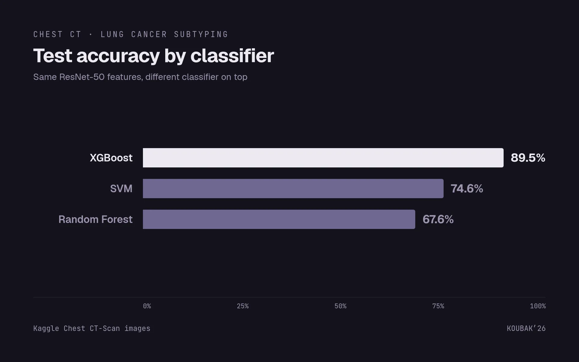
| XGBoost | 89.5% |
| SVM | 74.6% |
| Random Forest | 67.6% |
XGBoost was both the most accurate and the most stable across runs. KNN at
k=1 was the other strong performer.
Two findings mattered more than the headline number:
Every model exceeded 98% sensitivity on the Normal class. Separating healthy from diseased is close to solved on this data — which means the accuracy figure is carried substantially by the easy case, and reporting it alone would overstate how useful the system is.
The real difficulty is adenocarcinoma versus squamous cell carcinoma. The confusion matrices concentrate their errors there, between two subtypes that are genuinely similar in appearance. That, not overall accuracy, is where a clinically useful system would have to improve.
What I would do differently
The gap between 98% on Normal and the adeno/squamous confusion suggests the frozen ResNet-50 features are good at “is there a lesion” and weaker at the fine-grained texture distinctions between subtypes. Fine-tuning the backbone on this dataset, rather than using it off the shelf, is the obvious next experiment — as is reporting per-class F1 instead of overall accuracy, since on this problem the average hides exactly the part that matters.